

ChampDx's proprietary InfectISH™ technology is a novel RNA in-situ hybridization (ISH) assay capable of identifying bacteria and fungal pathogens down to the genus and species level while still preserving the morphological context.
Comprehensive Targets for Better Diagnosis
Our comprehensive target list for infectious disease takes the guess work out of identifying the correct pathogens associated with disease for veterinary laboratories.
Whether you are trying to identify a pathogen at the domain, kingdom, genus or species level our growing catalog of target probes are an invaluable tool for identifying probable pathogens at the single molecule resolution using only a standard brightfield microscope.
Our ever-growing catalog includes viruses, bacteria, dermatophytes, non-dermatophyte molds and yeasts.
InfectISH™ Workflow For Fungal Testing
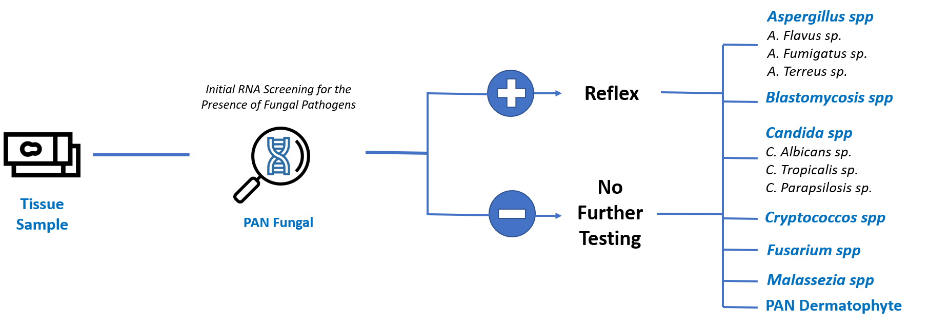
For veterinary diagnostic laboratory testing InfectISH™ is designed to first screen suspected tissues samples for the presence of fungal pathogens. If the fungal pathogen cannot be sufficiently identified for an appropriate treatment plan based on morphological analysis additional target probes are available for genus and species identification.
Because samples may be negative or morphological analysis alone is sufficient for optimum treatment selection, InfectISH™ saves time and cost for diagnostic laboratories, veterinarians and pet owners by avoiding unnecessary testing.
With just three tests fungal pathogens can be identified down to the species level!
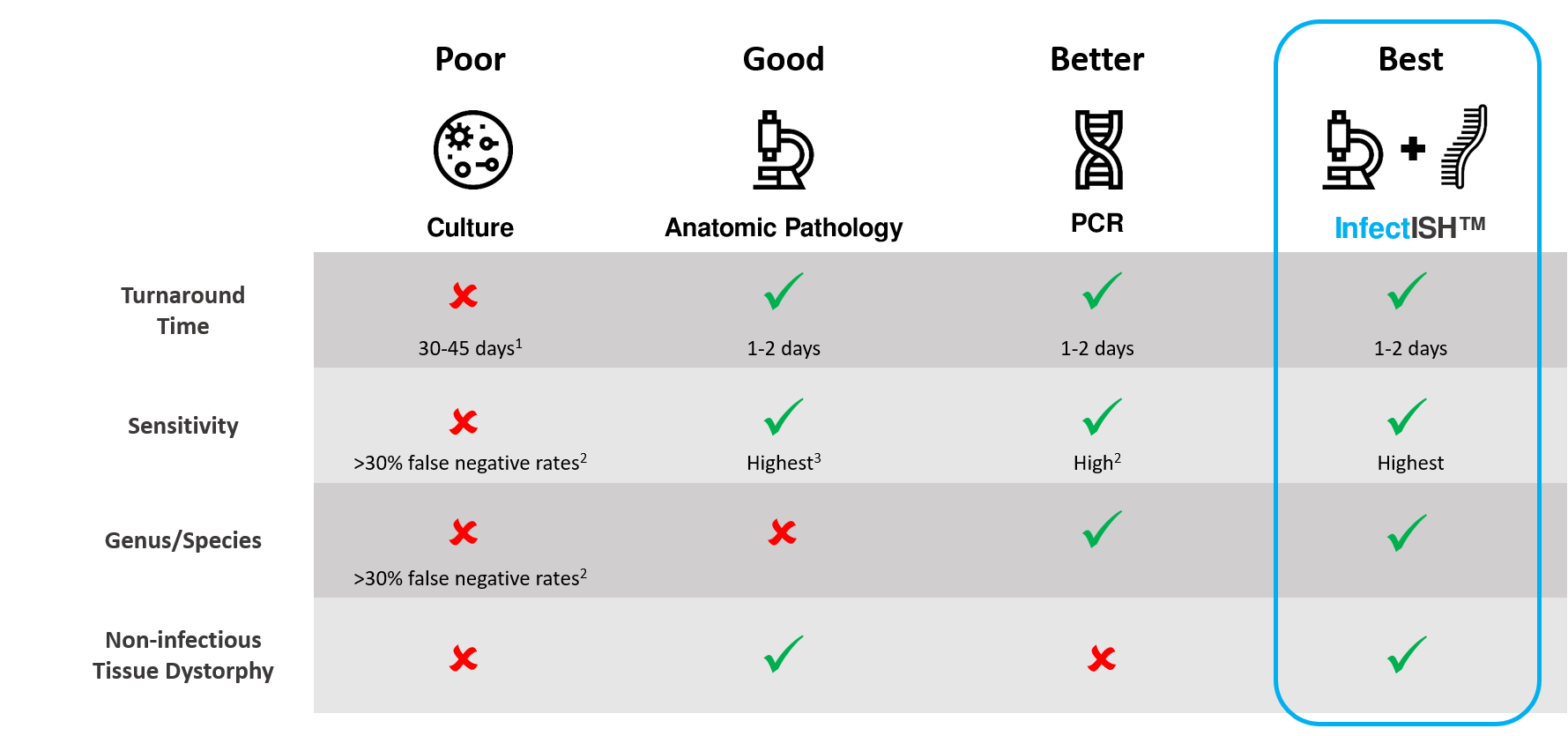
InfectISH™ combines the sensitivity of Anatomic Pathology with the specificity of PCR into a single Test
Culture: A gold Standard with Major Drawbacks
Culture has historically been the gold standard for fungal diseases like onychomycosis, but it is hindered by two big drawbacks: long turnaround times and poor sensitivity.
Fungal cultures routinely take between 2 to 4 weeks for results and even then many fungi are not able to be cultured in laboratories while others require biosafety level 3 precautions.
Cultures also suffer from poor sensitivity with ranges reported to be between 30% to 57% (2 and 4). This is epseicially problematic for dermatophytes, the leading cause of onychomycosis, due to their slow growth which can be inhibited by other saprophytic molds.
Anatomic Pathology: Sensitive but Poor Resolution
Special stains like periodic acid-Schiff (PAS) and Gomori methenamine silver (GMS) allow for the direct visualization of fungal elements like spores and hyphae, the degree of tissue involvement and the identification of other potential causes of disease.
Although PAS and GMS are faster and much more sensitive than culture they are unable to distinguish between different genera and species due to subtle nuances in fungal morphology limiting their use for targeted therapy.
An example of this is in the case of Cryptococcus which can have varying amounts of polysaccharide capsules that can make it difficult to distinguish from other probable fungi like Blastomyces, Candida and Histoplasma (5).
PCR and Sequencing: Resolution at the Cost of Accuracy
PCR and sequencing based assays have proven to be a powerful tool in identifying relevant infectious pathogens in a variety tissue samples. Unlike with culturing and and anatomic pathology PCR is capable of identifying the genus/species of the pathogen with specificity as high as 99.9%.
However, these grind and bind methods do not preserve the morphological context or measure the expression of the pathogen across the tissue. As a result PCR is prone to false-negative errors with a reported sensitivity ranging between 53.8% and 86% making it necessary to combine with PAS or GMS special stains to be clinically viable (2 and 6).
Furthermore, when PCR involves sequencing, tests can take up to 21 days consuming valuable time before a targeted therapy can chosen (6).
1. Bosshard PP. Incubation of fungal cultures: how long is long enough? Mucoses. 2011;54(5):e539-545. [PMID 21605185].
2. Gustafson E, Batkotic W, Bennett L, Page L, McCarthy L. DNA-based detection for onychomycosis correlates better to histopathology than does fungal culture. Dermatol Online J. 2019;25(7):1303/qt5bc2z46g. [PMID 31450272].
3. Sangoi AR, Rogers WM, Longacre TA, et al. Challenges and pitfalls of morphologic identification of fungal infections in histologic and cytologic specimens: a ten-year retrospective review at a single institution. Am J Clin Pathol. 2009;131(3):364-375. [PMID 19228642].
4. Petinataud D, Berger S, Ferdynus C, et al. Optimizing the diagnostic strategy for onychomycosis from sample collection to FUNGAL identification evaluation of a diagnostic kit for real-time PCR. Mycoses. 2016;59:304-311. [PMID 26806228].
5. Gazzoni AF, Severo CB, Salles EF, et al. Histopathology, serology and cultures in the diagnosis of cyptococcosis. Rev Inst Med Trop Sao Paulo. 2009;51(5):255-259. [PMID 19893977].
6. Meason-Smith C, Edwars EE, Older CE, et al. Panfungal Polymerase Chain Reaction for Identification of Fungal Pathogens in Formalin-Fixed Animal TIssues. Vet Pathol. 20017;54(4):640-648. [PMID 28346123]